
Health & Therapies
Equine Gastric Ulcers: Signs, Causes, and Definitive Diagnosis
Equine Gastric Ulcer Syndrome (EGUS) affects many horses, often without clear symptoms. Learn about the role of feeding and stress, and why only gastroscopy provides a definitive diagnosis.
Equidara editorial team · 27 September 2026 · 3 min read
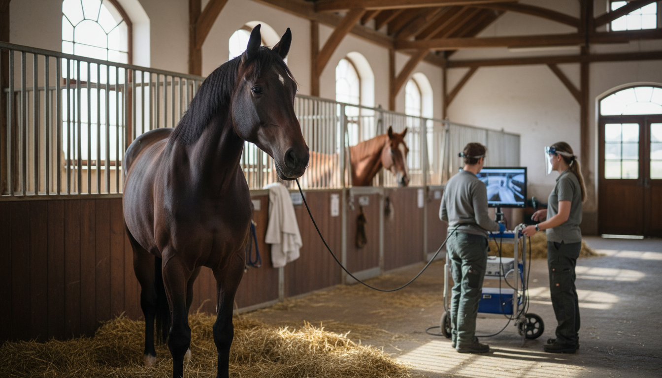
Gastric ulcers are prevalent in horses. Experts refer to this condition as Equine Gastric Ulcer Syndrome (EGUS). Performance horses and racehorses are particularly affected, with an incidence rate of up to 90 percent. Up to 50 percent of foals can also be affected. The signs are often vague, and the causes are diverse. Only gastroscopy provides a definitive diagnosis.
What is EGUS?
EGUS refers to gastric mucosal lesions that can occur throughout the stomach and adjacent small intestine. Two main forms are distinguished: lesions in the squamous portion (ESGD) and those in the glandular mucosa (EGGD). Both can occur simultaneously.
Signs: Often Non-Specific
The symptoms are rarely clear-cut. Many horses show only subtle changes that are easily overlooked. Typical signs include:
- Performance decline or reluctance under saddle
- Recurrent mild colic
- Poor appetite, weight loss, or a dull coat
- Behavioral changes such as yawning, flehmen, or increased recumbency
- Fecal water syndrome
There is no strong correlation between the severity of the ulcers and the clarity of the symptoms. Therefore, if you suspect ulcers, a veterinarian should be consulted.
Causes: Feeding, Stress, and Management
The development of gastric ulcers is multifactorial. Three key areas are important.
Feeding
Grain-rich diets with insufficient forage promote EGUS. The fermentation of grains produces volatile fatty acids (VFAs) that can damage the squamous mucosa of the stomach. Long periods without feed and insufficient hay exacerbate the problem because the stomach is then unprotected from acid. A study by FU Berlin showed that feeding barley significantly increased the risk of squamous lesions (ESGD), while alfalfa was associated with more glandular lesions (EGGD).
Conversely, slow eating has a beneficial effect. Hay nets can extend feeding time, thereby increasing saliva production. Saliva contains bicarbonate, which buffers stomach acid. In the Berlin study, horses that received hay from a net predominantly had no or only minor gastric findings.
Stress and Training
Stress is a significant risk factor. This includes intense training, competition, transport, frequent rider changes, or stable changes. Illnesses or hospital stays can also trigger gastric ulcers through stress mechanisms.
Management
The housing system influences the risk. Horses kept in year-round open stables with group contact and pasture access had significantly fewer gastric lesions in the Berlin study. In contrast, glandular lesions (EGGD) occurred significantly more often in horses kept in individual stalls with a paddock or in stalls with group pasture access.
Diagnosis: Only Gastroscopy Provides Certainty
Clinical signs and laboratory values alone are not sufficient for a definitive diagnosis. The only conclusive proof is achieved with a gastroscopy. For this procedure, an approximately three-meter-long endoscope is inserted through the nostril into the empty stomach.
For the veterinarian to fully assess the mucosa, the horse must be fasted for at least twelve hours. Water should always be available. Many horses require a muzzle during this time to prevent them from ingesting bedding.
During the examination, the lesions are graded according to a standardized scheme from 0 (normal) to 4 (severe). This allows for consistent communication and follow-up examinations.
If endoscopy is not available, the veterinarian may initiate a trial treatment. If symptoms improve within a few days with medication that reduces stomach acid, the diagnosis is considered probable. Which preparation is suitable and for how long it should be administered is solely decided by the veterinarian.
Common questions
What signs indicate gastric ulcers?
Typical signs are non-specific symptoms such as performance decline, recurrent mild colic, poor appetite, weight loss, flehmen, or fecal water syndrome. Since these signs can also occur with other conditions, veterinary clarification is necessary.
What are the most common causes of EGUS?
Grain-rich feeding, long periods without feed, insufficient forage, stress from training, transport, or stable changes, as well as individual stall housing without sufficient contact with other horses and pasture access, are among the most important risk factors.
How is a gastroscopy performed?
The horse must be fasted for at least twelve hours. Then, the veterinarian inserts an endoscope through the nose into the stomach and assesses the mucosa. The examination is the only definitive way to detect gastric ulcers and determine their severity.
Can gastric ulcers be treated without endoscopy?
If gastroscopy is not possible, the veterinarian may begin a presumptive treatment with an acid-reducing medication. If symptoms improve within a few days, it suggests EGUS. However, the precise diagnosis and therapy should always be managed by a veterinarian.
Sources
- Gastric Ulcers in Horses – Merck Veterinary Manual
- Magengeschwüre – Universitäres Tierspital Zürich
- Magengeschwüre – Universitäres Tierspital Zürich (Kurzinfo)
- Deutschlandweite multizentrische Untersuchung zur Ätiologie von Magenschleimhautläsionen beim Pferd – FU Berlin (2021)
- Prävalenz und Behandlungsmöglichkeiten von Läsionen der Drüsenschleimhaut beim Pferd – Pferdeheilkunde (2021)
- Magengeschwür beim Pferd
Written by Equidara editorial team. Last checked: 4 October 2026.
- Linseed and Linseed Mash for Horses: How to Properly Prepare This Traditional Home Remedy — Linseed mash is a time-honored home remedy for digestion and skin health. Here, you'll find preparation instructions from the University of Zurich, the daily dosage for your horse, and important considerations.
- How to Identify Good Hay: Smell, Color, Dust, Mold, and Hay Analysis — You can identify good hay by its color, smell, and texture – but only a lab analysis can confirm whether it's truly healthy for your horse. Here's how to check quality step by step.
- PPID in the Senior Horse: Recognizing Signs, the ACTH Test, and Daily Care — PPID (Equine Cushing's Disease) primarily affects horses over 15 years old. Learn how to recognize the signs, how your vet uses the ACTH test for diagnosis, and what you can do in daily life to help.
- Strangles in horses: how it spreads, the signs and what buyers should know — Strangles is a highly contagious infection of the upper airways. We explain the signs, how it spreads and what silent carriers are, and show how to bring a new horse onto your yard safely.
Related
- Services and jobs — Farrier, vet, training, horse share.
- Saddles and accessories — For the horse: saddles, rugs, bridles, hoof protection.